-
 Medical Gas Pipeline System (MGPS) in Bangladesh.
-
Operation Theatre (OT) Equipment
-
Critical Care (ICU & NICU)Equipment's
-
Infection Control & Consumable device
-
Medical Teaching Model Products
-
Hospital Curtain & Furniture
-
Diagnostics & Lab Equipment
-
Home used/Personal Healthcare
-
Medical Accessories & Consumables
-
Surgical Instrument
-
Refurbished Machine
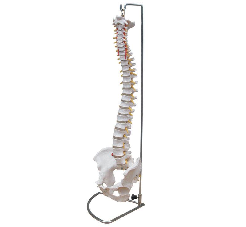
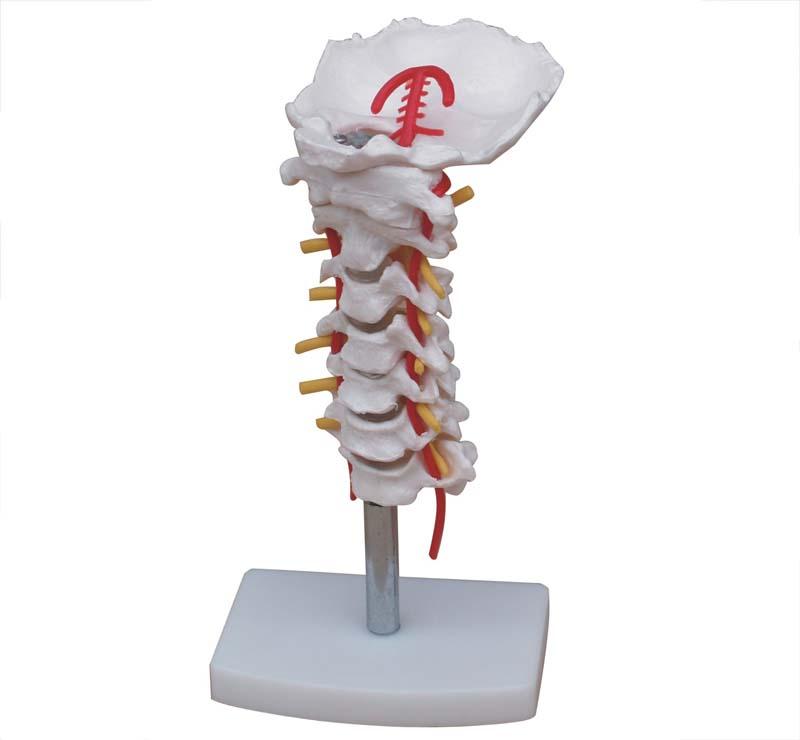
Vertebral Column with Pelvis
Inhouse product
-
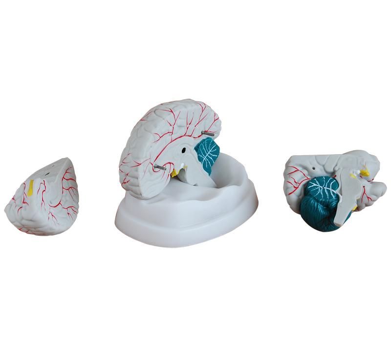
New Style Brain Model
৳10.00 -
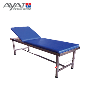
Patient Examination bed Model AHS-111
৳4,500.00 -
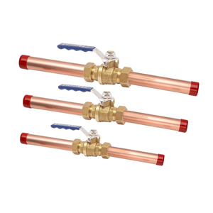
Line Valve, KITZ-Japan
৳3,499.00 -
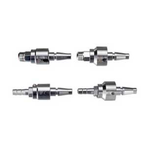
Oxygen ,AIR, Vac Probe
৳1,500.00 -
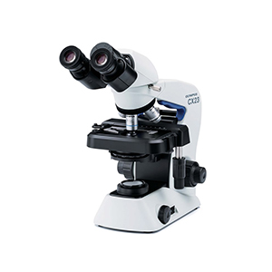
Olympus Microscope CX23 LED
৳165,000.00
- Product Name: Life-Size Vertebral Column with Pelvis
- Product No: XC-105
- Material: PVC
Note: we have attached all vertebral column ,if need details please download pdf file .
Description:
This model shows all significant features of each vertebra, including spinal cord, nerve roots, the vertebral artery, a herniated disc and vertebral notch etc. Special features include: flexible 29" tall vertebral column complete with pelvis, sacrum, occipital bone, vertebral artery, all nerve branches and herniated lumbar disc. Deluxe chrome stands 34" high.
Packing: 2pcs/carton, 80x32x39cm, 9kgs
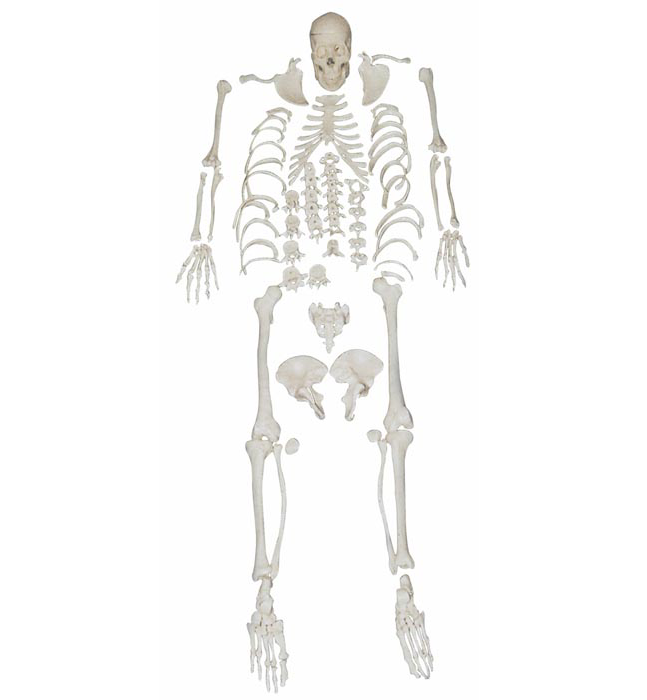
The human spine is an essential part of our body that provides support, protection, and flexibility. It consists of 33 individual vertebrae, each separated by intervertebral discs that act as shock absorbers. The vertebral column also includes the sacrum and coccyx, which form the pelvis.
Understanding the anatomy of the vertebral column and pelvis is crucial for medical professionals such as doctors, physical therapists, and chiropractors. It is also important for educators teaching anatomy and physiology to students. The "Vertebral Column with Pelvis" model is a valuable tool that accurately depicts the structure of the spine and pelvis.
The model is often used in medical schools, hospitals, and clinics to teach students and healthcare professionals about the different regions of the spine, their functions, and the intervertebral discs that provide cushioning between the vertebrae. It is also used to demonstrate the different curves of the spine, such as the cervical, thoracic, and lumbar curves, which are important for maintaining balance and stability.
The "Vertebral Column with Pelvis" model is typically made from materials such as plastic or resin, which are durable and easy to clean. The model is designed to be anatomically accurate, with each vertebra and intervertebral disc in the correct position and proportion. The model also includes the sacrum and coccyx, which are part of the pelvis.
In addition to being used for educational purposes, the "Vertebral Column with Pelvis" model is also used in clinical settings to diagnose and treat conditions related to the spine and pelvis. For example, it can be used to identify the location of spinal cord injuries or herniated discs. It can also be used to demonstrate proper posture and body mechanics, which can help prevent back pain and other spinal disorders.
When examining the model, it is important to understand the different regions of the spine and their associated functions. The cervical spine, which is located in the neck, is responsible for supporting the head and allowing for movement in multiple directions. The thoracic spine, which is located in the upper back, provides stability for the ribcage and protects the internal organs. The lumbar spine, which is located in the lower back, is responsible for supporting the body's weight and allowing for movement in multiple directions.
The intervertebral discs that separate each vertebra act as shock absorbers and help prevent the vertebrae from rubbing against each other. The discs are composed of a tough outer layer called the annulus fibrosus and a soft inner layer called the nucleus pulposus. When a disc herniates, the inner layer can push through the outer layer, causing pain and inflammation.
The sacrum and coccyx are part of the pelvis, which is responsible for supporting the weight of the upper body and providing attachment points for muscles and ligaments. The pelvis is also important for childbirth, as the baby must pass through the pelvic opening during delivery.
In conclusion, the "Vertebral Column with Pelvis" model is an essential tool for medical professionals and educators in teaching and understanding the anatomy of the human spine and pelvis. Its accurate depiction of the different regions of the spine and their associated functions makes it a valuable resource in diagnosing and treating spinal conditions, as well as preventing them through proper posture and body mechanics.
Related products

Giant Ear Model
Giant Ear Model
New Style Brain Model
Giant Ear Model
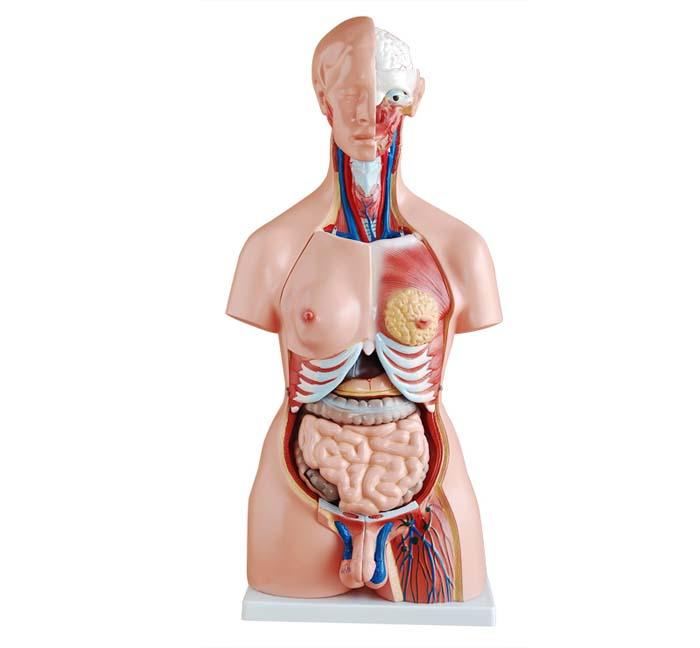

Male Torso 13 Parts 42CM
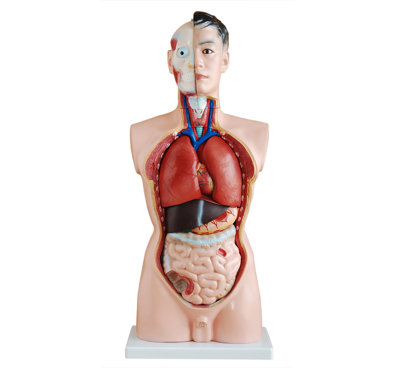
85CM Male Torso 19 Parts

Contact Info
- Address: House : J7-B. Extensions Pallabi ,Rupnagar ,Mirpur-11, Dhaka-1216 Bangladesh Tel: +8802 55013276 Email: info@ayatht.com, info.ayatht@gmail.com WeChat & WhatsApp No: +8801913587092
- Phone: +880 1719 351 166
- Email: info.ayatht@gmail.com
My Account
Medical Equipment Suppliers in Bangladesh - Ayatht
The page and briefly explain the importance of reliable medical equipment suppliers in the healthcare industry. Get access to the top medical equipment list in Bangladesh for all your healthcare needs. Our comprehensive list of devices and equipment ensures you have everything you need to provide the best possible care. Contact us today to learn more. (Medical device suppliers) Welcome to our Ayatht Medical Equipment Suppliers page, where we provide you with the best medical supply companies and distributors in the industry. We understand the crucial role that medical equipment plays in the healthcare sector, and we aim to connect you with reliable suppliers that offer high-quality products and excellent customer service. our source for the most comprehensive medical equipment list in Bangladesh. Our company offers a wide range of medical equipment and supplies from the best suppliers in the industry.
Our Medical Equipment Suppliers Services:
We provide a wide range of medical supplies, including surgical instruments, diagnostic equipment, medical consumables, and much more. List the types of medical equipment and supplies that are available from the suppliers listed on the page. (Medical equipment manufacturers) - (Medical equipment distributors) At Aytatht, we understand how crucial it is for medical professionals to have access to high-quality medical equipment. That's why we are proud to offer a comprehensive list of medical equipment in Bangladesh, making it easier for healthcare providers to find the equipment they need to provide the best possible care for their patients. Our listed medical equipment suppliers offer a wide range of products, including but not limited to surgical instruments, diagnostic equipment, hospital furniture, medical disposables, and more. With our selection of top-rated suppliers, you can find everything you need to equip your healthcare facility or organization. (Medical equipment wholesale suppliers)
How it Works:
Explain how the process of finding and purchasing medical equipment from the suppliers listed on the page works. At Ayatht, we strive to make the process of acquiring and maintaining medical equipment in Bangladesh as simple and straightforward as possible. (Healthcare equipment suppliers) Our process is simple and straightforward. You can browse our selection of top-rated medical equipment suppliers to find the products you need. Once you've found the right supplier, you can contact them directly to request a quote or place an order. With our easy-to-use platform, finding and purchasing medical equipment has never been easier. We begin by understanding your specific needs and requirements, and then we source the equipment you need from our network of top suppliers. We then provide installation and maintenance services, ensuring that your equipment is always in top condition. Ordering medical supplies from our medical suppliers in Dhaka is easy. Browse our website or call us to select the products you need, and we will deliver them to your doorstep.
Our Medical Equipment List in Bangladesh Services:
At Ayatht, we offer a range of medical equipment services to meet the diverse needs of healthcare providers in Bangladesh. Our services include equipment procurement, maintenance, repair, and replacement. (Medical supply companies) Our medical equipment suppliers are carefully selected to offer you the best possible experience. We prioritize quality products, fair prices, fast shipping, and excellent customer service. With our suppliers, you can trust that you'll receive the best possible equipment at a price you can afford.
Process & Methodology:
Explain the process and methodology used to select the medical equipment suppliers listed on the page. Our process and methodology are designed to ensure that we provide the highest quality medical equipment and services to healthcare providers in Bangladesh. Our process and methodology include understanding your specific needs, sourcing equipment from top suppliers, providing installation and maintenance services, and offering equipment replacement when necessary. We are committed to providing the best possible medical equipment and services to healthcare providers in Bangladesh. (Medical instrument suppliers) We use a rigorous selection process to ensure that our listed medical equipment suppliers meet our high standards. Our team carefully evaluates each supplier based on factors such as quality of products, pricing, shipping options, and customer service. Only the best suppliers make it onto our list, so you can trust that you're getting the best possible equipment and service.
Problem Solved:
Explain how the medical equipment suppliers listed on the page can help solve the problem of finding reliable sources for medical equipment. At Ayatht, we understand the challenges healthcare providers face when it comes to acquiring and maintaining high-quality medical equipment. That's why we offer comprehensive medical equipment services in Bangladesh to help solve these problems.(Hospital equipment suppliers) Finding reliable medical equipment suppliers can be a challenge, especially when it comes to quality products, fair prices, and excellent customer service. Our listed suppliers are carefully chosen to ensure that you have access to a selection of the best medical equipment and supplies on the market. With our trusted suppliers, you can be confident that you're getting top-quality products that meet your needs also include equipment procurement from top suppliers, equipment maintenance and repair, and equipment replacement when necessary. We provide a one-stop-shop solution for all your medical equipment needs. Our services are designed to help healthcare providers in Bangladesh overcome the challenges of acquiring and maintaining high-quality medical equipment. With our comprehensive list of medical equipment and services, we aim to make it easier for healthcare providers to focus on providing the best possible care for their patients. Address the common challenges faced by customers when looking for medical suppliers in Dhaka. Finding a reliable supplier who provides top-quality products at affordable prices can be challenging. Our medical suppliers in Dhaka solve this problem by offering a vast selection of products at competitive rates.
Key Features:
List the key features of the medical equipment suppliers listed on the page, such as quality products, affordable prices, fast shipping, and excellent customer service.(Medical equipment dealers) Wide range of medical equipment available Equipment procurement from top suppliers Equipment maintenance and repair services Equipment replacement when necessary Our medical suppliers in Dhaka offer 24/7 customer support, timely delivery, competitive pricing, and a vast selection of high-quality medical supplies.
Use Cases:
Provide examples of the types of organizations or businesses that can benefit from the medical equipment suppliers listed on the page. Our medical equipment services are designed to meet the diverse needs of healthcare providers in Bangladesh, including hospitals, clinics, and other healthcare facilities. Our medical suppliers in Dhaka have provided medical supplies to hospitals, clinics, and medical professionals across the city. By choosing our medical suppliers in Dhaka, you can save time and money, access a vast selection of high-quality medical supplies, and enjoy excellent customer service. CTA: Contact us today to request a quote or learn more about our services!